Science Team Press Contact Request Deck

ULTRA-EARLY CANCER DETECTION

Your cells emit light.
We learned to read it.

Detects cancer 100-200× earlier than standard imaging.
No blood draw. No biopsy. No radiation.

Cancer is caught too late.

Every 60 seconds, the U.S. loses one person to cancer. Not because treatments fail, but because they start too late.

>90%
5-year survival
when caught early
~30%
5-year survival
when caught late

The problem isn't timing. It's what we can see.

A single cell acquires a driver mutation.
Atypical daughter cells multiply.
A pre-malignant cluster forms. Pre-cancer
A diffusion-limited microtumor takes shape. Carcinoma in situ
Hypoxia triggers angiogenesis. New blood vessels feed the growth. Carcinoma in situ
The tumor breaches the basement membrane and invades surrounding tissue. Stage I
Standard imaging typically detects a tumor at ~1 cm. Roughly 1 billion cells. By then, it's been growing silently for years.

But there's a signal before there's a tumor.

The Science

Cancer has a light signature.

Every cell in your body emits faint light as a byproduct of metabolism. Cancer cells process energy differently, and the light they produce shifts: altered wavelengths, changed intensity. This happens at the metabolic level, before any mass forms.

Dr. Nirosha Murugan spent a decade building technology that reads these signals with enough precision to identify cancer at 100-200x fewer cells than standard imaging.

Normal cells emit a smooth, steady light signature. Cancer cells emit a disrupted one.
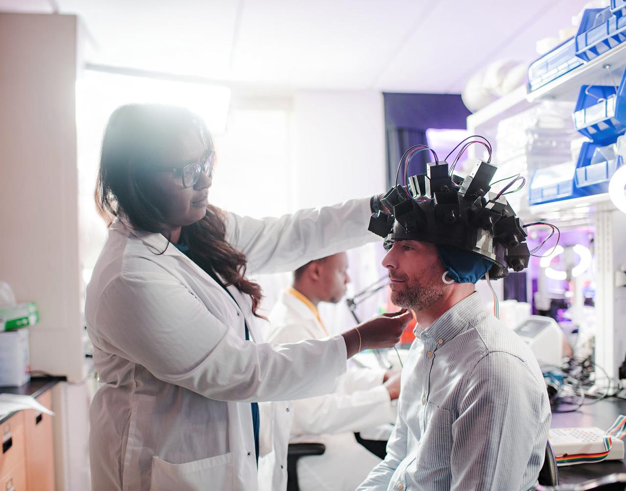
The Scan

We built the tool to read it.

We've built prototype devices and validated them in peer-reviewed studies. Now we're developing the clinical version.

A patient sits in a dark room for about fifteen minutes. A sensor reads the faint light coming off their body. AI analyzes the signal. Same visit, same day.

No blood draw. No biopsy. No radiation.

Two systems. One signal.

QSense

Hardware

QSense™

A sensor array sensitive enough to count individual photons from living tissue. Non-invasive, radiation-free, and designed to detect cancer signals before a tumor forms.

Functional prototype, ready for human studies

LuminAI

Software

LuminAI™

AI trained on a decade of cell and animal data. Processes roughly a million data points per scan, distinguishing healthy tissue from cancer in real time.

Fully developed, integrated with hardware

"These sensors were built to detect faint light from distant stars. We pointed them at the faint light coming off the human body, and built an AI that uses it to read the state of your cells and tell healthy tissue from cancer."

Dr. Nirosha Murugan, Interim CEO, Chief Scientist & Co-Founder

Explore the published research →
Dr. Nirosha Murugan in the laboratory

What It Can Detect

Proven in the lab.
Heading to humans.

HelioFlux skin cancer detection device render
Featured · Human Study In Preparation

Skin Cancer
Detection

90-92% accuracy across multiple cancer cell lines in cell cultures and animal studies. Detected cancer within 24 hours of exposure. Tumors weren't physically detectable for another 18 days. First human study targeting Q3 2026 with a Beverly Hills dermatology partner.

Dr. Murugan fitting photon-sensing helmet on research subject
Published · 2025 · Cell Press

Brain
Health

World's first study reading brain activity through light alone. Published in iScience (Cell Press), 2025. Tracked brain activity in 20 living subjects, cross-validated against EEG. The same platform is designed to monitor brain health after chemotherapy, concussion, and neurodegeneration.

Mitochondria cellular metabolism visualization
Research Frontier

Metabolic &
Systemic Disease

The metabolic disruptions in cancer also appear in diabetes, cardiovascular disease, and autoimmune conditions. Published research shows light tracks those disruptions directly. HelioFlux is pursuing that. Cancer is first, but the platform is designed to go further.

Read metabolism through light. Cancer is just the beginning.

The scientist behind
the technology.

Dr. Nirosha Murugan
Dr. Nirosha Murugan
Interim CEO, Chief Scientist & Co-Founder

Canada Research Chair in Tissue Biophysics. Trained in Michael Levin's lab at Tufts and the Harvard Wyss Institute. 40+ peer-reviewed publications. $1.6M+ in active research grants. She built this science. HelioFlux is how it reaches the world.

Meet the full team →

Featured In

Scientific American NPR Radiolab Cell Press CBC News LA Business Journal
Read press coverage →

NVIDIA Inception Program Member

The science works.
The next step is human data.

Ten years of published research. Validated in cells, animals, and living human subjects. We're raising a pre-pilot round for our first IRB-approved human study.

Request Investor Deck Schedule a Conversation →
© 2026 HelioFlux Inc. All rights reserved. HelioFlux is in research-stage validation. Not FDA cleared. Not intended to diagnose or treat any disease.
¹ Imaging detectability is often discussed around ~1 cm3 (~1 g), roughly ~108–109 cells (Frangioni JV, 2008; Davis L et al., Cancers 2022).
² Tumor growth modeling shows time to detectability can range from ~2 years (fast) to ~10 years (moderate), depending on doubling time (Holland et al., NCBI Bookshelf).